
 Türkçe


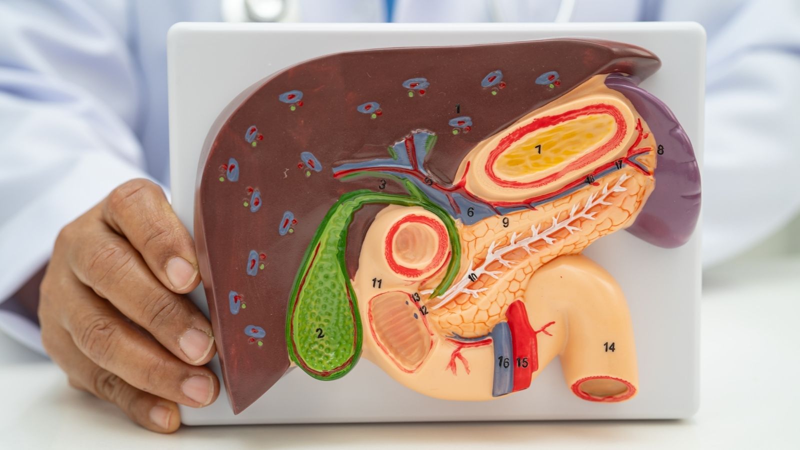
Hepatopancreatobiliary Surgery is a highly specialized subspecialty of General Surgery applied in the treatment of benign and malignant diseases of the liver, pancreas, and bile ducts. This discipline encompasses a wide and complex spectrum ranging from major cancer surgery operations such as the Whipple procedure to liver transplantation. In treatment, in addition to open methods, the most up-to-date technologies such as minimally invasive and robotic surgery, which offer the patient faster recovery, are utilized. Success requires an experienced surgical team with mastery of the anatomy of these delicate organs and advanced technological equipment.

Bile Duct Surgery
Gallbladder Surgery

Whipple Procedure

Liver Surgery

Liver Cyst Surgery
You can contact Prof. Dr. Ebru Soy, one of the doctors performing Hepatopancreatobiliary Surgery in Ankara, to get information about Hepatopancreatobiliary Surgery prices, the process, and the hospital where Hepatopancreatobiliary Surgery is performed.

Prof. Dr. Ebru Ayvazoğlu Soy
Education;
High School: TED Ankara College – Graduation: 1998
Faculty of Medicine: Hacettepe University Faculty of Medicine – Graduation: 2005
Specialization: General Surgery Residency Training – Başkent University Faculty of Medicine, Department of General Surgery – 2012
Post-Specialization Career;
2012–2014: Compulsory public service – Muş State Hospital
January 2014 – November 2022: Başkent University Faculty of Medicine, Department of General Surgery – Served as a specialist physician and later as an academician
March 2016 – October 2017: Başkent University Istanbul Practice Hospital – Deputy Director Responsible for Liver Transplantation
June 2020 – November 2022: Başkent University Ankara Hospital – Director of the International Patient Department
Why Is an Army of Experts Required for Successful Hepatopancreatobiliary Surgery?
In today’s medical understanding, especially in the treatment of serious and complex diseases such as cancer, progressing with the decisions of a single physician is now a thing of the past. The secret to achieving the best outcomes in hepatopancreatobiliary (HPB) surgery lies in the “multidisciplinary team” approach, where specialists from different medical branches come together. We can liken this approach to a sports team in which each player is the best in their position and determines a common strategy to win the match. The goal is to evaluate the patient’s condition from every aspect and draw the most accurate, safest, and personalized treatment roadmap.
This process is carried out through regular meetings called the “tumor board.” In these meetings, each patient is discussed in detail with their unique condition. This expert team generally includes the following:
- HPB Surgeon
- Medical Oncologist
- Radiation Oncologist
- Radiologist
- Interventional Radiologist
- Gastroenterologist
- Pathologist
- Specialist Nurses
- Dietitians
- Shutterstock
Thanks to this collective intelligence, vital questions such as “Is surgery the most appropriate treatment for this patient?”, “Is pre-treatment such as chemotherapy needed before surgery?”, and “What should the timing of the surgery be?” are answered in the most accurate way. It should not be forgotten that the risk of complications after these surgeries is higher compared to other operations. This team not only makes the surgical decision but also anticipates potential problems that may arise in the postoperative period, takes precautions, and intervenes rapidly when necessary. In short, success in HPB surgery is measured not only by the skill at the tip of the scalpel but also by the strength of the large team behind the hand holding that scalpel.
Which Factories of the Body Does Hepatopancreatobiliary Surgery Deal With?
This surgical branch deals with a group of organs located in the upper part of our abdomen, each undertaking vital functions in its own field. Let us take a closer look at these organs and the diseases we frequently encounter.
Liver: The largest internal organ of our body and virtually a chemical factory. It cleans our blood of toxic substances, produces bile for digestion, synthesizes proteins, and stores energy. In this organ with so many functions, conditions requiring surgery are also quite diverse. The main diseases addressed by liver surgery are:
- Primary liver cancers (those developing due to Hepatitis B and C)
- Cancers spreading to the liver from other organs such as the colon (metastases)
- Benign tumors that grow excessively and cause problems
- Liver cysts
Pancreas: A leaf-shaped organ hidden just behind our stomach. It has two basic functions: first, to produce digestive enzymes to break down the food we eat; second, to secrete the insulin hormone that balances our blood sugar. Pancreatic diseases often progress insidiously and their surgery is quite difficult. The main diseases addressed by pancreatic surgery are:
- Pancreatic cancer (the most common and most dangerous)
- Neuroendocrine tumors of the pancreas
- Cystic tumors with malignant potential
- Severe chronic pancreatitis attacks
Gallbladder and Bile Ducts: A system consisting of a small sac that stores the bile produced by the liver and thin channels that carry this bile to the duodenum for digestion. Both benign and malignant diseases can be seen in this system. The main diseases addressed by biliary system surgery are:
- Gallbladder cancer
- Bile duct cancer (cholangiocarcinoma)
- Complex gallstone diseases
- Bile duct injuries occurring during surgeries
- Strictures in the bile ducts
What Is the Golden Rule of Defeating Cancer with Hepatopancreatobiliary Surgery?
In cancers developing in HPB organs, surgical treatment has a single main objective: to provide complete cure for the patient. The most fundamental and indispensable principle on the path to this goal is called “R0 resection.” This technical term actually has a very simple meaning: complete removal of the tumor together with a margin of healthy tissue around it, leaving not a single cancer cell behind. We can liken this to pulling out a wild weed in the garden together with its root, ensuring that no piece remains under the soil. If even the smallest part of the root remains, there is a risk that the weed will grow again. The situation is the same in cancer surgery. When the removed tissue is examined by the pathologist, it must be confirmed that the cut margins do not contain cancer cells. This “clear surgical margin” is the most important determinant for the patient to live a long and healthy life.
There are two other important elements that complement this golden rule. The first is lymph node dissection. Cancer cells generally use the lymphatic pathways like a highway to spread in the body. Therefore, during surgery, not only the tumor but also the surrounding lymph nodes connected to it are removed. This procedure both eliminates potential sites of spread and allows us to determine the true stage of the cancer, helping us decide whether additional postoperative treatment (such as chemotherapy) is necessary.
The second is accurate staging. Sometimes insidious tumors such as pancreatic and gallbladder cancers may have spread into the abdomen in tiny particles too small to be seen on imaging. To understand this situation, a procedure called “staging laparoscopy” is sometimes performed before the main surgery. By entering the abdomen with a small camera, the peritoneum and the surface of the liver are examined. If such spread is detected, we protect the patient from the risks of a very major and non-beneficial surgery and direct them straight to more effective chemotherapy treatment.
To Make an Appointment for Diseases in the Field of General Surgery
You can contact us via WhatsApp by clicking the button.


Which Strategies Are Applied During Liver Surgery?
One of the most admirable features of the liver is its ability to regenerate at an extraordinary speed. This feature allows surgeons to safely remove up to 70% of the liver. The main surgical approaches for liver tumors are as follows:
Liver Resection (Hepatectomy): This is the most commonly applied method and involves removal of the tumorous part of the liver. The surgeon’s choice is shaped according to the location and size of the tumor and the health status of the patient’s liver. There are basically two different philosophies. The first is anatomical resection; here, one or more of the liver segments separated according to its own vascular structure are removed as a whole. The second is parenchyma-sparing resection; here, without adhering to anatomical boundaries, only the tumor and a margin of healthy tissue around it are removed, preserving the maximum amount of healthy liver. Especially in patients with an underlying disease such as cirrhosis, this second method is more frequently preferred to prevent the remaining liver from going into failure.
Liver Transplantation: It is the most definitive treatment method for liver cancers (HCC) developing on the basis of cirrhosis. Because transplantation eliminates not only the cancerous tissue but also the entire diseased liver that has the potential to continuously produce new cancer. However, due to the limited availability of organ donation, only patients whose tumor number and size fall within certain criteria (for example, the Milan Criteria) can benefit from this treatment opportunity.
Tumor Ablation (Burning) Treatments: This is a method applied in cases where surgical resection is risky or when tumors are small and few in number. The aim is to eliminate the tumor in its place without surgery. Different types of energy are used for this purpose. The main ablation treatments applied are:
- Radiofrequency Ablation (RFA)
- Microwave Ablation (MWA)
- Irreversible Electroporation (IRE) – Non-thermal ablation
What Is the Process of the Whipple Procedure, the Most Challenging Pancreatic Surgery?
Pancreatic surgeries are considered the pinnacle of HPB surgery due to their technical complexity and high risks. The standard operation applied for tumors located in the head of the pancreas is “pancreaticoduodenectomy,” commonly known as the Whipple procedure. This operation is a very major and demanding process that involves the removal of many interconnected organs like a domino effect, followed by reconstruction of the digestive system.
In the first stage of the surgery, all adjacent tissues with the potential for cancer spread are removed as a whole. The structures removed during the Whipple procedure are:
- The head of the pancreas
- The entire duodenum
- A portion of the stomach
- The gallbladder
- The lower end of the main bile duct
- Surrounding lymph nodes
The second and most delicate stage of the surgery is reconstruction, that is, rebuilding. After the organs are removed, the remaining organ ends are reconnected to the small intestine to ensure continuity of the digestive system. This is like installing a new digestive plumbing system. In sequence, the remaining pancreas, the main bile duct, and the stomach are sutured to the small intestine. The most critical and leakage-prone of these connections is the one between the pancreas and the intestine. Aggressive pancreatic enzymes leaking from this suture line can lead to serious infections and bleeding within the abdomen. Therefore, the success of the surgery depends not only on clean removal of the tumor but also on the flawless construction of these connections. For tumors located in the body and tail of the pancreas, distal pancreatectomy is performed. In this surgery, if the tumor is benign, preservation of the spleen is of great importance for the patient’s immune system.
How Is Surgical Treatment Planned in Gallbladder and Bile Duct Cancers?
In cancers of this region, the treatment strategy is determined according to the exact millimetric location of the tumor. This requires surgical planning with the precision of a watchmaker.
- Gallbladder Cancer: A painful reality of these cancers is that most of them are incidentally discovered in the pathology laboratory after a routine gallbladder surgery performed due to gallstone complaints. If the cancer is at a very early stage (involving only the innermost layer), the first surgery is sufficient. However, if the cancer has progressed a little deeper into the wall of the gallbladder, then the first surgery is insufficient and the patient needs a second, more extensive operation. In this “radical” surgery, a portion of liver tissue from the bed where the gallbladder is attached and the lymph nodes in the region are removed.
- Bile Duct Cancer (Cholangiocarcinoma): This cancer is divided into three different types depending on where it originates in the bile ducts, and each has a completely different operation. The main types planned according to its location are:
- Intrahepatic: The tumor is in the ducts within the liver. Its treatment is liver surgery (hepatectomy).
- Hilar Region (Klatskin Tumor): The tumor is at a critical junction where the right and left main bile ducts coming out of the liver meet. Its treatment is a very major and technically challenging operation in which the bile ducts, a large portion of the liver, and the lymph nodes are removed together.
- Distal: The tumor is at the lower end of the bile duct passing through the pancreas. Therefore, its treatment is the Whipple procedure, as in pancreatic head cancer.
When the bile duct is removed in these surgeries, to ensure the flow of bile, one end of the small intestine is sutured to the bile duct in the liver to create a new pathway called “Roux-en-Y.”
Contact Us.
You can request an online appointment by filling out the form. Our team will contact you as soon as possible.
What Advantages Do Minimally Invasive Methods Offer to Patients in Hepatopancreatobiliary Surgery?
While in traditional open surgery the abdomen is opened with a large incision from top to bottom, minimally invasive surgery (also known among the public as closed or bloodless surgery) is performed with a camera and special instruments advanced through a few small incisions. The two main types of this technology are laparoscopy and robotic surgery. Initially used for simpler operations, these methods are now successfully applied even in the most complex operations of HPB surgery in high-volume, specialized centers thanks to increasing experience and advancing technology.
In robotic surgery, the surgeon sits at a console and controls robotic arms that precisely mimic their hand movements under a three-dimensional and magnified view. This provides precision and range of motion that the human hand cannot achieve, especially in narrow and deep areas. The tangible benefits offered to patients by these modern approaches are quite numerous. The main advantages offered by minimally invasive methods are:
- Less postoperative pain
- Less blood loss
- Smaller surgical scars
- Reduced risk of infection
- Shorter hospital stay
- Faster return to daily life and work
- Opportunity to start postoperative chemotherapy earlier
It has been scientifically proven that robotic surgery significantly increases spleen preservation rates, especially in surgeries of the pancreatic tail. However, these advanced technology surgeries require a serious training process and high experience. Therefore, performing such complex minimally invasive surgeries only in centers specifically focused on this field and by experienced surgeons is essential for patient safety.
What Are the Possible Complications of These Major Surgical Operations?
Hepatopancreatobiliary surgery inherently involves serious risks and potential complications. A modern HPB center stands out not only with its ability to perform flawless surgery but also with its capacity to anticipate, prevent, and effectively manage these possible problems. Close postoperative monitoring of the patient is at least as critical as the surgery itself. The main complications that may be encountered are:
- Pancreatic Fistula (Leakage): It is the most feared condition after pancreatic surgeries. Digestive enzymes leaking from the cut end of the pancreas or the suture line can cause serious damage, abscess, and bleeding within the abdomen.
- Liver Failure: Especially after major liver surgery, it is the inability of the remaining liver tissue to meet the body’s needs. To minimize this risk, very detailed planning is performed before surgery.
- Bile Leak: Leakage of bile into the abdomen from the cut surface of the liver or the bile duct connection. It can usually be managed without surgery, through endoscopic methods (ERCP).
- Delayed Gastric Emptying: Especially after the Whipple procedure, it is the slowing of stomach movements and inability to empty normally. Although it temporarily complicates the patient’s nutrition, it usually improves over time.
- Bleeding: A serious risk that may originate from major vessels in the surgical area.
- Infection: Development of an abscess at the surgical site or within the abdomen.
Management of these complications requires a multidisciplinary approach. In most cases, instead of a second surgery, minimally invasive interventions performed by interventional radiologists or gastroenterologists (such as drain placement or stent insertion) are preferred to resolve the problem.
Are There Innovations That Accelerate the Recovery Process in Hepatopancreatobiliary Surgery?
Yes, absolutely. Modern approaches in HPB surgery focus not only on techniques in the operating room but also on improving the entire process from the moment the patient steps into the hospital until the day of discharge. The most important innovations in this field are as follows:
Enhanced Recovery After Surgery (ERAS) Protocols: This is an evidence-based care package aimed at ensuring that the patient is affected as little as possible by the physiological stress created by surgery and returns to normal life as soon as possible. It has replaced traditional, memorized practices. The main components of the ERAS protocol are:
- Elimination of prolonged preoperative fasting
- Preparation of the patient with special nutritional solutions before surgery
- Avoiding unnecessary fluid overload during surgery
- Use of modern, multimodal methods for pain control
- Mobilizing and walking the patient immediately after surgery
- Transitioning to oral nutrition as soon as possible
Neoadjuvant Therapy (Preoperative Treatment): This strategy reverses the treatment sequence, especially in some aggressive tumors such as pancreatic cancer. Instead of performing surgery first and then giving chemotherapy, chemotherapy and/or radiotherapy are applied first, followed by planned surgery. There are many logical reasons for this approach: it shrinks the tumor, allowing surgery to be performed more easily and more cleanly; it targets microscopic cancer cells that may have spread throughout the body but are not yet visible; and most importantly, it protects patients with aggressive tumor biology that does not respond to treatment from the burden of major surgery.
Advanced Imaging Technologies During Surgery: These are the surgeon’s greatest assistants during the operation. Intraoperative Ultrasound (IOUS) is like a “GPS device” in the surgeon’s hand. By placing it directly on the organ, it allows real-time visualization of the exact location of the tumor and its relationship with vessels. Indocyanine Green (ICG) Fluorescence Imaging is based on the principle of a special dye given intravenously glowing under infrared light. Thanks to this technology, the bile ducts become visible like a roadmap, small metastases in the liver that cannot be seen with the naked eye can be detected, or the boundaries of the liver segment to be removed can be clearly delineated. These technologies greatly contribute to making surgeries safer and more effective.