
 Türkçe


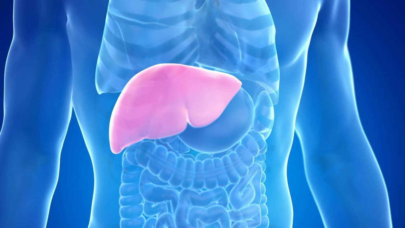
Hepatectomy, also known as liver resection surgery, is the procedure of surgically removing a tumorous or diseased portion of the liver. This procedure is of vital importance in the treatment of masses in the liver. The most fundamental factor that makes the operation possible is the liver’s extraordinary capacity for self-renewal (regeneration), unlike any other organ in the body. The main goal of surgery is to remove the diseased area while ensuring that a sufficient amount of healthy liver tissue remains so that the body can fully perform its functions. This delicate balance is the key to the success of modern liver surgery.
You can contact Prof. Dr. Ebru Soy, one of the doctors performing Hepatectomy (Liver Resection) Surgery in Ankara, to get information about Hepatectomy (Liver Resection) Surgery prices, the process, and the hospital where Hepatectomy (Liver Resection) Surgery is performed.

Prof. Dr. Ebru Ayvazoğlu Soy
Education;
High School: TED Ankara College – Graduation: 1998
Faculty of Medicine: Hacettepe University Faculty of Medicine – Graduation: 2005
Specialization: General Surgery Residency Training – Başkent University Faculty of Medicine, Department of General Surgery – 2012
Post-Specialization Career;
2012–2014: Compulsory public service – Muş State Hospital
January 2014 – November 2022: Başkent University Faculty of Medicine, Department of General Surgery – Served as a specialist physician and later as an academician
March 2016 – October 2017: Başkent University Istanbul Practice Hospital – Deputy Director Responsible for Liver Transplantation
June 2020 – November 2022: Başkent University Ankara Hospital – Director of the International Patient Department
What Is Hepatectomy (Liver Resection)?
Hepatectomy is the surgical removal of all or part of the liver. It is generally performed due to liver tumors, metastases, benign masses, or trauma. It is divided into two types: partial (parenchymal) and total. This procedure is planned with the liver’s regenerative capacity in mind and generally aims to preserve the healthy portion of the liver. The treatment process requires a multidisciplinary approach.
Why is planning liver surgery so important?
The success of liver surgeries depends not so much on the operating room as on meticulous preoperative planning. Our biggest goal is to avoid the serious problem we call postoperative “liver failure” (PHLF). This condition means that the remaining liver cannot meet the body’s needs and, unfortunately, it is the most serious risk factor.
Therefore, a standard risk assessment is essential for every patient. This assessment becomes even more critical especially in patients with an underlying liver disease (such as Hepatitis B, cirrhosis) or those who have received intensive chemotherapy. Because long-term chemotherapy can impair the quality of healthy liver tissue by causing a condition called “chemotherapy-associated liver injury” (CALI).
When evaluating, we look not only at the condition of the liver but also at the patient’s overall health resilience. Factors such as nutritional status, muscle mass (sarcopenia or the presence of “muscle wasting”), and overall frailty directly affect how well the body can withstand this major surgical stress.
Which tests are used to measure liver strength (reserve)?
To understand whether the liver can tolerate surgery, we need to know its “functional reserve,” i.e., its working capacity. Some chronic disease scores used in the past (such as the MELD or Child-Pugh score) are insufficient in this regard. These scores show the general chronic state of the liver but cannot predict how well it will handle the sudden and severe metabolic stress created by surgery.
In surgical planning, we rely on “dynamic” tests. The ‘gold standard’ in this field is the ICG (Indocyanine Green) test. In this test, a special green dye (ICG) is administered intravenously. Only the liver clears this dye from the body. After 15 minutes, we check how much of this dye remains in the blood (ICG-R15).
If the liver is working quickly and efficiently, it clears the dye immediately (low R15). This is a good sign for surgery. But if the liver is working slowly, most of the dye remains in the blood (high R15). This indicates that the liver reserve is low and that there is a high risk. This dynamic test provides us with a much more accurate and valuable insight into the liver’s real-time working capacity than other scores.
How is the volume of the remaining liver (FLR) calculated?
The second pillar of planning is calculating the volume of the liver we will leave behind. We call this the “Future Liver Remnant” (FLR). Based on CT or MRI images, we calculate the volume of this portion in cubic centimeters and relate it to the total liver volume (FLRV%).
As a general rule, an FLR of 20–25% may be sufficient in a healthy liver. However, things are not always that simple.
If the patient’s liver is already damaged due to cirrhosis, hepatitis, or chemotherapy injury (CALI), the quality of that liver is reduced. In this case, even a volume of 30%, or even 40%, may be functionally insufficient. In other words, the issue is not only how much volume we leave behind, but how well that volume works. Making a decision based solely on volume would dangerously overlook the risk of liver failure in these patients.
Is there a special method (functional volumetry) that measures both volume and quality?
Yes—this is where the most advanced tools of modern medicine come into play: Functional Volumetry.
In this method, we combine two different types of data:
- Volume (Volumetry): The FLR volume we obtain with CT/MRI.
- Function (Scintigraphy): A special nuclear medicine test called Hepatobiliary Scintigraphy (HBS) (performed with 99mTc-mebrofenin).
HBS shows us, as a percentage, how well different regions of the liver function and how function is distributed within the liver. For example, HBS may tell us that the portion we plan to leave behind (FLR) accounts for 50% of total liver function, while the portion we will remove accounts for the other 50%.
We then combine these two data points: we multiply the FLR volume (FLRV%) by the functional percentage of that region. This yields a clear, individualized, and highly sensitive result regarding the true working capacity of the liver that will remain after surgery. This integrated approach provides much greater safety—especially in risky or damaged livers—than relying on volume alone.
What are the differences between these various tests that assess liver reserve?
The assessment methods and their roles in surgical planning are as follows:
- Child-Pugh Score: Determines the overall severity of cirrhosis. It is weak in predicting surgical risk.
- MELD Score: Evaluates end-stage chronic liver disease and kidney function. It is insufficient in predicting surgical stress.
- ICG-R15 (Indocyanine Green): Measures how quickly the liver clears the dye from the blood. It is the preoperative ‘gold standard’ functional test.
- Volumetry (CT/MRI): Determines the anatomic volume of the remaining liver (FLR). It is a basic requirement.
- Functional Volumetry (with HBS): Combines volume (Volumetry) and function (HBS). It provides the most sensitive risk prediction, especially in damaged livers.
To Make an Appointment for Diseases in the Field of General Surgery
You can contact us via WhatsApp by clicking the button.


What is the treatment strategy when colon cancer spreads to the liver (metastasis)?
When colon cancer spreads to the liver (stage 4 cancer), treatment requires a multidisciplinary approach; that is, the surgeon, medical oncologist, and radiologist work as a team. In these patients, the most important treatment that offers a chance of long-term survival is the surgical removal of these liver metastases.
Sometimes, when patients present, the tumors in the liver may not be suitable for surgery. In this case, a strategy called “conversion therapy” is followed. The patient is first started on neoadjuvant chemotherapy (NCT). The purpose of this chemotherapy is to shrink the tumors and make them operable.
However, there is a very delicate balance here: the duration of chemotherapy. Studies show that more than 5–6 cycles of chemotherapy, while not significantly increasing tumor response, increases the risk of damaging healthy liver tissue (CALI). This, in turn, raises the risk of postoperative liver failure.
What is the difference between ‘anatomic’ and ‘non-anatomic’ resection in metastasis treatment?
We can think of the liver as an orange consisting of 8 segments. Each segment (slice) has its own blood vessel and bile duct:
- Anatomic Resection (AR): In this method, the entire segment or sector (several segments) containing the tumor is removed by following the blood vessels of that region.
- Non-Anatomic Resection (NAR or Wedge Resection): Also known as “parenchyma-sparing surgery.” Here we do not look at segment boundaries. We remove only the tumor itself and a small amount of healthy liver tissue around it (enough to leave a safe margin), essentially “scooping it out.”
In metastatic disease (CRLM), the main goal is to remove the tumor with an R0 margin, leaving no cancer cells behind. Studies have shown that as long as this R0 margin is achieved, Non-Anatomic Resection (NAR) provides oncologic outcomes at least as good as anatomic resection.
The greatest advantage of NAR is that it preserves much more healthy liver tissue. This is vital in two ways: first, the patient needs reserve to tolerate postoperative chemotherapy. Second, if the disease recurs in the future, there will be enough liver left for a chance at repeat surgery.
Is the same surgery (NAR) applied for primary liver cancer (HCC or ICC) as well?
Generally, no. These cancers tend to spread along the vascular pathways within the segment. Therefore, especially at certain stages, Anatomic Resection (AR) has been associated with better long-term survival outcomes in terms of complete cancer clearance. Unlike metastasis, in these tumors, removing not only the tumor but also the anatomic pathway (segment) along which it can spread may be oncologically superior.
Why is a biopsy not performed for liver lesions found incidentally during surgery?
This is a very important multidisciplinary rule. If, for example, while a patient is undergoing bowel (colon) surgery, the surgeon sees a suspicious small lesion on the liver, they should absolutely not biopsy it. A biopsy performed at that moment can cause tumor cells to spread into the abdominal cavity (“seeding”). This can negatively affect the success of the planned definitive liver surgery and the patient’s long-term survival. The patient should first recover from the bowel surgery, undergo detailed imaging, and then have a planned liver operation.
What is done if the remaining liver volume (FLR) is not large enough?
Sometimes, we calculate that the remaining liver (FLR) after removing the tumor will be too small to be sufficient for the patient. At this point, we surgically trigger the liver’s miraculous ability to regenerate. If we block blood flow to the diseased portion we plan to remove, the body directs all blood to the healthy portion, and that healthy portion (FLR) begins to grow rapidly.
How do the PVE and ALPPS methods work to enlarge the liver?
There are basically two methods to achieve this growth:
Portal Vein Embolization (PVE): This is a non-surgical procedure performed by interventional radiology. Through an angiographic approach, the main blood vessel (portal vein) that supplies the diseased liver portion to be removed is blocked with special materials. Blood flow is redirected to the healthy side, and that side begins to grow. This growth usually takes 4 to 8 weeks. Its disadvantage is the risk that the tumor may continue to grow during this waiting period.
ALPPS (Two-Stage Surgery): This is a newer and more aggressive surgical technique.
- Stage Surgery: The surgeon performs the first operation. First, the healthy liver and the diseased liver tissue are physically separated (partition). Then, the portal vein branch going to the diseased side is tied off (ligation). This dual intervention triggers very rapid growth (hypertrophy) in the healthy side (FLR).
- Stage Surgery: About 1–2 weeks later, when the FLR reaches an adequate size, the patient is taken to surgery again and the non-functioning diseased liver portion is completely removed.
ALPPS provides much faster growth than PVE. However, the classic ALPPS procedure has a much higher risk, complication rate, and mortality compared to PVE. Therefore, modified techniques such as “partial-ALPPS” or “mini-ALPPS,” which significantly reduce risks, have now been developed.
How are the risks of ALPPS surgery managed?
Because ALPPS is a high-risk procedure, patient selection is extremely strict. The ALPPS Risk Score is used to predict the risk of a “futile outcome” (i.e., the patient being lost before reaching the second stage or shortly after completing it).
This scoring looks not only at risk factors before Stage 1 but primarily at the physiologic response after Stage 1. If the patient’s bilirubin or creatinine (kidney function) values rise after the Stage 1 operation, or if a serious complication develops, this is the most important indication that the body cannot tolerate this stress. These findings show that the likelihood of a fatal outcome is very high if the patient proceeds to Stage 2 and may lead the surgeon to abandon performing Stage 2.
Contact Us.
You can request an online appointment by filling out the form. Our team will contact you as soon as possible.
Can liver surgery be performed with a minimally invasive (laparoscopic or robotic) method?
Yes. In recent years, liver surgery has experienced a major shift from open surgery toward Minimally Invasive Liver Surgery (MILS). This means laparoscopic (LLR) or robotic (RLR) surgery.
- Minor Resections: For small tumors located in “favorable” segments of the liver (anterior and inferior parts; 2, 3, 4b, 5, 6), laparoscopic surgery is now accepted as a standard and safe option in experienced centers.
- Major or Difficult Resections: Removal of more than 3 segments (major hepatectomies) or tumors located in “technically difficult” segments (superior and posterior parts; 1, 4a, 7, 8) are highly complex operations. Such surgeries should be performed only in specialized “centers of excellence.”
What are the advantages of robotic surgery compared to laparoscopic surgery?
Both methods can yield excellent results in expert hands. However, robotic surgery (RLR) has a clear advantage, especially in those hard-to-reach, deep, posterior, and superior segments (1, 4a, 7, 8):
The robot’s arms can rotate 720 degrees at angles that the human wrist or straight laparoscopic instruments cannot achieve, and they can operate without tremor. This provides the surgeon with tremendous technical superiority in those challenging regions. Robotic operations performed in these regions have been reported to be associated with less blood loss and a lower need to convert to open surgery (“conversion”) compared to laparoscopy. Therefore, the robotic platform is increasingly preferred for the highest-complexity minimally invasive liver resections.
How are bleeding control and liver transection performed during surgery?
Various techniques are used during surgery to control bleeding and transect the liver safely:
- Pringle Maneuver: Temporary and intermittent clamping of the main blood vessels supplying the liver.
- Clamp-Crush Method: Still the most common and safest technique for transecting liver tissue. It gently separates the tissue, allowing vessels and bile ducts to be seen and controlled.
- Kellyclasia (Robotic Surgery): The robotic counterpart of the ‘clamp-crush’ technique performed with robotic arms.
- Vessel Sealers/Staplers: Used to seal large vessels and ducts.
Is there a special surgical maneuver to prevent liver failure in cirrhotic patients?
Yes—especially in cirrhotic and high-risk patients (with a high ICG-R15 value), there is a very subtle technique applied when performing minimally invasive surgery:
The liver has a ligament called the “Round Ligament” (RL) or “Ligamentum Teres.” In cirrhotic patients, this ligament can reopen and act as a “safety valve” that decompresses high blood pressure within the liver (portal hypertension).
In minimally invasive (MILS) surgeries, this ligament is usually cut to create working space. However, studies have shown that preserving the Round Ligament in these high-risk cirrhotic patients significantly reduces the rates of severe postoperative liver failure and ascites (fluid accumulation in the abdomen). This is a very important physiologic protective maneuver aimed at reducing risk in this delicate patient group.
What navigation technologies guide the surgeon during surgery?
We no longer perform liver surgeries solely with anatomical knowledge and tactile feedback. Advanced imaging technology guides us.
- 3D Planning and Virtual Surgery: Before surgery, the patient’s CT images are converted into a 3D model with specialized software. The surgeon can virtually simulate the operation on this model.
- ICG Fluorescence Imaging: During surgery, ICG (Indocyanine Green) dye is administered to the patient again. When viewed with a special (infrared) camera, ICG makes liver tissue, tumors, or bile ducts “glow” bright green. This helps detect small tumors not visible to the naked eye or identify bile leaks.
- Augmented Reality (AR): Especially in robotic surgery, the preoperatively prepared 3D virtual model can be projected onto the real-time camera view during the operation. This allows the surgeon to operate as if “seeing inside” the liver and to know where deep vessels not visible on the surface are located.
What do ERAS protocols include to accelerate postoperative recovery?
In postoperative care, we apply modern protocols called Enhanced Recovery After Surgery (ERAS) Programs. The aim is to reduce the stress the body experiences from surgery and accelerate liver regeneration.
The main components of these programs are:
- Effective pain control that avoids strong narcotics
- Getting out of bed as soon as possible (early mobilization)
- Starting oral nutrition as soon as possible
- High-protein dietary recommendations
- Ensuring adequate fluid intake
One of the most important warnings: strictly avoiding alcohol for the first six weeks after surgery so the liver can complete its repair process.
How is postoperative liver failure (PHLF) defined and what are its grades?
Postoperative liver failure (PHLF) is monitored worldwide using a standard definition and grading system (ISGLS criteria). This is important so that we can compare outcomes objectively. This definition is based on elevated bilirubin and impaired coagulation (INR) values measured on or after postoperative day 5.
This grading is directly related to the patient’s vital risk:
- Grade A PHLF: Mild, transient impairment. No change in patient management is required.
- Grade B PHLF: Worsens the patient’s course. Requires non-invasive interventions (such as medication or fluid adjustment).
- Grade C PHLF: Severe condition progressing to multiple organ failure. Requires intensive care (ICU) and invasive treatments (dialysis, ventilator).
The purpose of all our preoperative planning (ICG, HBS, FLR volume) is to protect the patient from Grade B and especially Grade C failure.
How is bile leak managed if it occurs after surgery?
Bile leak is a known risk especially after complex and long liver surgeries. After high-risk procedures, a drain may be placed in the abdomen to monitor this condition.
If a bile leak is confirmed, treatment is generally first to drain (evacuate) the bile in that area and start antibiotics to prevent infection. If the leak does not stop with simple methods and especially if it originates from the main bile ducts (“central type”), then a second surgical intervention (bilioenteric anastomosis—i.e., connecting the bile duct to the intestine) may be required to repair the leak.
Frequently Asked Questions
How Long Does Hepatectomy (Liver Resection) Surgery Take?
Hepatectomy is the surgical removal of part of the liver and usually takes between 3 and 6 hours. The duration may vary depending on the size of the liver segment to be removed, the location of the tumor or lesion, and the patient’s overall health status.
Which Doctor Should You See for Hepatectomy (Liver Resection)?
The specialty to consult for this procedure is general surgery, particularly surgeons specialized in hepatopancreatobiliary surgery.