
 Türkçe


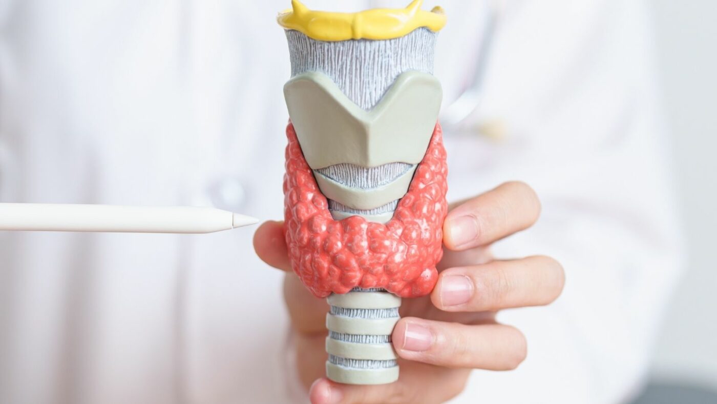
Parathyroid adenoma surgery, medically called parathyroidectomy, is the surgical removal of a benign parathyroid tumor (adenoma) that is the source of excessive parathyroid hormone (PTH) production and disrupts the body’s calcium balance. This operation eliminates the underlying cause of dangerously elevated blood calcium levels (hypercalcemia). The surgery is typically performed through the front of the neck, targeting this small but overactive gland, and it is the only definitive curative treatment aimed at permanently correcting the disturbed calcium metabolism.
You can contact Prof. Dr. Ebru Soy, one of the doctors performing Parathyroid Adenoma Surgery in Ankara, to get information about Parathyroid Adenoma Surgery prices, the process, and the hospital where Parathyroid Adenoma Surgery is performed.

Prof. Dr. Ebru Ayvazoğlu Soy
Education;
High School: TED Ankara College – Graduation: 1998
Faculty of Medicine: Hacettepe University Faculty of Medicine – Graduation: 2005
Specialization: General Surgery Residency Training – Başkent University Faculty of Medicine, Department of General Surgery – 2012
Post-Specialization Career;
2012–2014: Compulsory public service – Muş State Hospital
January 2014 – November 2022: Başkent University Faculty of Medicine, Department of General Surgery – Served as a specialist physician and later as an academician
March 2016 – October 2017: Başkent University Istanbul Practice Hospital – Deputy Director Responsible for Liver Transplantation
June 2020 – November 2022: Başkent University Ankara Hospital – Director of the International Patient Department
What Is Primary Hyperparathyroidism (PHPT)?
Primary hyperparathyroidism (PHPT) is a condition characterized by excessive production of parathyroid hormone (PTH) by the parathyroid glands. This leads to elevated blood calcium levels. The most common cause is a parathyroid adenoma. PHPT can present with symptoms such as bone loss, kidney stones, fatigue, and depression. Diagnosis is made by measuring blood calcium and PTH levels. Treatment is usually surgical parathyroidectomy.
How is hyperparathyroidism diagnosed and what are its subtypes?
The diagnosis is primarily established through blood tests. The most important finding we look for is persistently elevated blood calcium (usually above 10.4 mg/dL). To ensure this elevation is not transient, it should be confirmed in at least two separate measurements.
The key diagnostic point is this: while calcium is high, the PTH level is also elevated at the same time (often >65 pg/mL). Normally, when calcium rises, the body’s protective mechanism should suppress PTH production—so PTH would be expected to be very low. In PHPT, however, the autonomous adenoma does not “hit the brakes” and continues producing hormone. This combination (high calcium + high PTH) clearly establishes PHPT.
Sometimes we encounter a subtype called “Normohormonal PHPT.” Here, blood calcium is still elevated, but PTH is not “overtly high”; instead it sits at the upper end of the normal range (for example, 40–65 pg/mL). This is still abnormal, because in the presence of high calcium, PTH should not be “normal” but rather “suppressed” (very low). Even a normal-range PTH in this context shows the gland is not responding appropriately to high calcium (autonomous function), and these patients also benefit from surgical treatment.
What are the symptoms of high calcium?
Elevated calcium levels can negatively affect many systems in the body. In medicine, this classic presentation is summarized as “bones, stones, abdominal groans, and psychic moans.” Major problems that can result from this condition include:
- Recurrent kidney stones
- Frequent urination
- Bone loss (osteoporosis)
- Bone and joint pain
- Easy fractures
- Chronic fatigue and weakness
- Muscle weakness
- Forgetfulness
- Difficulty concentrating
- Depressive mood or anxiety
- Stomach pain
- Ulcers
- Nausea and vomiting
- Constipation
- Pancreatitis
- High blood pressure (hypertension)
- Heart rhythm problems
Of course, not all symptoms have to occur together. In fact, a substantial proportion of patients today are asymptomatic and are diagnosed incidentally after hypercalcemia is detected on routine blood tests performed for another reason.
Who needs parathyroidectomy, and when is it necessary?
Surgery is the only permanent and definitive (curative) treatment for PHPT. Some medications, such as “cinacalcet,” can temporarily lower PTH secretion and therefore blood calcium by increasing the parathyroid gland’s sensitivity to calcium. However, these medications are not a “cure”; they manage the biochemical problem without removing the adenoma and must typically be used long-term.
International clinical guidelines strongly recommend surgery in certain situations. Indications for surgery include:
- Blood calcium levels significantly above the upper limit of normal
- A history of kidney stones
- Very high urinary calcium excretion
- Reduced kidney function (decreased GFR)
- Osteoporosis or osteopenia on bone density testing (DEXA)
- Age under 50 years
- Symptoms listed above (fatigue, bone pain, etc.)
- Choosing surgery knowingly even if asymptomatic
Especially in patients under 50, surgery is recommended because the disease persisting for many years carries a high risk of permanent damage to bones, kidneys, and the cardiovascular system.
Why are preoperative imaging and a “road map” so important?
The foundation of modern parathyroid surgery is preoperative localization imaging. In the past, surgeons operated without knowing where the adenoma was and had to open both sides of the neck (Bilateral Neck Exploration – BNE) to find and inspect all four parathyroid glands one by one.
Today, the goal for most patients is minimally invasive parathyroidectomy (MIP). The prerequisite for MIP is clearly “marking” the location of the diseased adenoma before surgery. Successful imaging provides the surgeon with a “road map,” allowing a focused approach through a small 1–2.5 cm incision directly over the abnormal gland.
This targeted approach significantly reduces hospital stay (often same-day discharge) and costs. If imaging does not show a clear location or gives conflicting results, the surgeon may need to revert to the traditional BNE method during the operation. Therefore, high-quality and accurate imaging is critical for the success of modern surgery.
To Make an Appointment for Diseases in the Field of General Surgery
You can contact us via WhatsApp by clicking the button.


Which standard imaging methods are used to locate the diseased gland?
As a first step, we typically use anatomical and functional imaging together. Standard methods include:
- High-resolution ultrasonography (US)
- Parathyroid scintigraphy (Sestamibi SPECT/CT)
Ultrasonography is excellent for mapping the anatomy of the neck. When performed by an experienced radiologist, it has a sensitivity of about 72% to 89% for detecting a single adenoma. It is also very valuable for planning where the surgeon will place the incision.
Parathyroid scintigraphy is a “functional” test. A radioactive tracer (Technetium-99m sestamibi) is injected intravenously. This tracer is taken up by a hyperfunctioning parathyroid adenoma, and on imaging the adenoma appears as a “bright” focus.
Using both methods (US and Sestamibi) together, with concordant findings (both pointing to the same location), can increase localization rates to as high as 96%.
Which advanced imaging tests are used when standard methods are insufficient?
Sometimes—especially when the gland is very small, located in an unusual place, or when multiple glands are diseased—standard imaging can be insufficient or produce conflicting results. In such cases, we turn to more advanced methods to obtain a clearer surgical road map. These include:
- 4-dimensional computed tomography (4D-CT)
- F-fluorocholine (FCH) PET/CT
4D-CT is a specialized dynamic contrast-enhanced CT technique that uses the “time” factor (the fourth dimension). Parathyroid adenomas show a characteristic pattern of contrast enhancement followed by rapid “washout” compared with thyroid tissue or lymph nodes. 4D-CT captures this pattern and demonstrates the adenoma’s location with excellent anatomical detail, reaching sensitivities up to 92.5% in single-gland disease.
FCH PET/CT is currently among the most sensitive functional imaging methods available. It is based on the principle that hyperactive parathyroid cells use “choline” at a high rate. It is particularly useful when other methods fail (persistent or recurrent disease) or when very small adenomas are being sought, with reported sensitivities approaching 99%.
What does “ectopic” (abnormally located) parathyroid gland mean?
During fetal development, the parathyroid glands normally “migrate” to their typical location behind the thyroid. Sometimes this journey ends in an atypical position, and the glands settle outside their usual anatomical site. This is called an “ectopic” parathyroid gland.
Ectopic adenomas are one of the most important reasons for failed initial surgery or disease recurrence. The surgeon may check the typical neck locations, but the adenoma is not there because it is “hidden” elsewhere. Ectopic adenomas are estimated to occur in about 14% of cases. Common ectopic locations include:
- The thymus (upper chest)
- The superior mediastinum
- Behind the esophagus
- Retropharyngeal space
- Inside the thyroid gland (intrathyroidal)
- Within the carotid sheath
That is why advanced imaging such as 4D-CT or PET—capable of surveying these “hidden” areas—and the surgeon’s mastery of anatomical variations are vital for surgical success.
What is minimally invasive parathyroidectomy (MIP), and what are its advantages?
Minimally Invasive Parathyroidectomy (MIP) is the preferred “gold standard” surgical method for the majority of PHPT patients today (about 80–85%). Its essential prerequisite is that preoperative imaging has clearly localized the diseased adenoma.
Instead of opening the entire neck, this technique uses a very small incision (typically 1 to 2.5 cm) on the side where the adenoma is located. The surgeon targets the abnormal gland directly, removes it while protecting the vocal nerve and the nearby normal parathyroid tissue, and completes the operation in a focused manner. Cure rates for MIP (about 98.6%–99.4%) are equal to or better than the traditional approach. Its advantages are clear:
- Much smaller scar (better cosmetic outcome)
- Less postoperative pain
- Significantly lower complication rates
- Shorter operative time
- Very short hospital stay (often same-day discharge after 4–6 hours of observation)
- Faster return to normal life and work
When is the traditional “bilateral neck exploration” (BNE) necessary?
Bilateral Neck Exploration (BNE) involves a wider midline incision (typically longer than 4 cm), mobilization of both sides of the thyroid region, and identification and visual evaluation of all four parathyroid glands. When performed by experienced surgeons, it remains a traditional method with high success rates (about 95–98%).
Despite the widespread adoption of MIP, BNE is still required in certain situations:
- Preoperative imaging (US, Sestamibi, 4D-CT) fails to clearly localize the adenoma
- Imaging results are discordant or conflicting
- Suspicion of multi-gland disease
- Hyperparathyroidism associated with familial/genetic syndromes (e.g., MEN)
- Need for simultaneous thyroid surgery (e.g., thyroidectomy)
- Failure to find the adenoma during an MIP approach or failure of rapid PTH testing (conversion from MIP to BNE)
Why is intraoperative “rapid PTH testing” (IOPTH) so vital?
This is one of the most important safety “locks” in modern parathyroid surgery. Intraoperative Parathyroid Hormone (IOPTH) testing measures the PTH level from blood samples taken immediately after the adenoma is removed.
The logic is simple: PTH has a very short biological half-life (about 3–5 minutes). If the surgeon successfully removes the main source of excess PTH, the PTH level should fall rapidly.
This test answers—while the patient is still asleep on the operating table—“Did you remove the correct gland?” and “Is there another diseased gland left behind?” The widely accepted confirmation approach is based on the “Modified Miami Criteria,” which requires two conditions to be met to confidently end the surgery. First, the PTH level should drop by at least 50% at 10–15 minutes compared with the highest pre-excision (baseline) value. But that alone is not enough. Second—and more importantly—the final PTH value must also fall definitively into the laboratory’s normal reference range.
When both conditions are met, the operation is considered successful with accuracy approaching 99%, allowing the surgeon—especially in MIP cases—to safely conclude without opening the other side of the neck unnecessarily.
Contact Us.
You can request an online appointment by filling out the form. Our team will contact you as soon as possible.
How are my vocal cords and healthy glands protected during surgery?
Parathyroid surgery has two core quality measures: removing the diseased gland (cure) and avoiding damage to healthy tissues—especially the vocal nerve and the remaining normal parathyroid glands (safety). Modern surgery uses advanced technologies to achieve this. These include:
- Intraoperative Neuromonitoring (IONM – Nerve Monitoring)
- Near-Infrared Fluorescence Imaging (NIRF/ICG)
Nerve monitoring (IONM) helps protect the recurrent laryngeal nerve (RLN), which controls vocal cord motion and lies very close to the parathyroid region. IONM assists the surgeon in identifying the nerve, confirming its anatomical course, and—most importantly—verifying that it remains functionally intact throughout the operation. This minimizes the risk of nerve injury and postoperative hoarseness.
Fluorescence imaging (NIRF/ICG) is used to help prevent permanent low calcium (hypoparathyroidism). Another surgical risk is compromised blood supply to the remaining healthy parathyroid glands or accidental removal. With the ICG technique, a special dye (Indocyanine Green) is injected intravenously during surgery, and well-perfused, healthy parathyroid glands glow brightly under a specialized camera. This allows the surgeon to confirm that the glands left behind are viable and well perfused, significantly reducing the risk of permanent hypoparathyroidism.
What is the success rate after parathyroidectomy, and can the disease recur?
Parathyroidectomy is a highly successful operation, especially when performed by experienced surgeons and centers. Long-term cure rates are routinely above 95%, and with focused MIP techniques they approach 99%. “Success” is defined as sustained normalization of blood calcium and PTH levels after surgery.
Recurrence rates are very low, generally below 2%. The benefits of successful surgery are often seen quickly; bone density begins to improve, and long-term (10-year) risk of major fractures decreases significantly.
Why does low calcium (hypocalcemia) happen after surgery?
In the first days after surgery, the most common issue is transient hypocalcemia. There are two main reasons:
First, the remaining healthy parathyroid glands may experience temporary “stunning.” After months or years of being suppressed by the dominant adenoma, these glands may pause function briefly due to surgical stress. It may take a few days for them to ramp up to full capacity again.
Second—and more importantly—there is a condition called “Hungry Bone Syndrome.” During the disease, high PTH levels continuously “leach” calcium from the bones into the bloodstream. The bones become “hungry” for calcium. Once the adenoma is removed and PTH drops abruptly, bone metabolism flips: the bones rapidly shift into a rebuilding mode and start absorbing calcium, phosphorus, and magnesium from the blood. This can cause deep and persistent drops in blood calcium levels.
What are the symptoms of low calcium?
It is very important for patients to recognize these symptoms and inform the care team if they occur. The most common early symptoms of hypocalcemia include:
- Numbness around the lips
- Numbness at the tip of the tongue
- Tingling in the fingertips
- Numbness in the hands and feet
- Muscle twitching
- Muscle cramps
- (In more severe cases) spasms in the hands and feet (tetany)
How are “Hungry Bone Syndrome” and hypocalcemia managed?
Because of the risk of postoperative hypocalcemia—especially the potential for Hungry Bone Syndrome—almost all patients are discharged with an oral calcium supplementation protocol.
However, in patients who develop Hungry Bone Syndrome or are at high risk (very high preoperative calcium and PTH), management must be more aggressive. The key point is that giving calcium alone is often not sufficient. This is because the “stunned” glands may also struggle to convert vitamin D into its active form (calcitriol). Without active vitamin D, intestinal absorption of calcium remains low.
Therefore, modern management aims to correct this mineral imbalance proactively. These patients typically receive both high-dose oral calcium (often 1 to 3 grams per day depending on severity) and active vitamin D (calcitriol) capsules together. Magnesium—critical for bone metabolism and PTH function—must also be monitored closely and supplemented orally or intravenously if needed. This intensive support continues until the bones become “repleted” and the healthy parathyroid glands resume full function, after which doses are gradually tapered based on weekly calcium checks.
What is the follow-up process after modern parathyroid surgery?
Modern parathyroid adenoma surgery is built on three pillars: excellent preoperative “targeting” (localization imaging), technology-supported “focused and safe surgery” (MIP, IOPTH, nerve monitoring, ICG), and careful “postoperative metabolic management” (calcium and vitamin D supplementation).
After surgery, once calcium levels normalize and stabilize, patients are typically followed to ensure durable cure with blood calcium and PTH checks at around 3 months, 6 months, and then annually.
Frequently Asked Questions
How Long Does Parathyroid Adenoma Surgery Take?
Parathyroid adenoma surgery usually takes 1 to 2 hours. The duration depends on how clearly the adenoma is localized, whether it is single-gland or multi-gland disease, and the chosen surgical technique (minimally invasive vs traditional).
Which Doctor Should You See for a Parathyroid Adenoma?
When a parathyroid adenoma is diagnosed, the appropriate specialist to consult is an endocrine surgeon. General surgeons specialized in endocrine surgery plan and perform surgical removal of the adenoma.